
Ovary
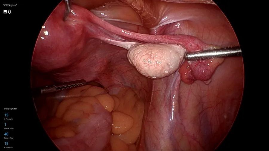
Ovarium sinistrum
- Paired structure: one on the left and one on the right side of the body.
The ovary (from Latin ōvārium 'egg') is the primary sex organ in the female reproductive system. It is the gonad in females responsible for the production and development of oocytes and their release as mature egg cells. The egg cells are also known as gametes, and the process of their development is gametogenesis, known as oogenesis in the female. The ovary also produces hormones to assist in the development of the egg cells. During their development each oocyte is contained in an ovarian follicle.
Many of the structural and functional features of the human ovary are shared with other mammals and vertebrates and ovaries of some kind are found in many invertebrates. This article is primarily about the human ovary.
The paired ovaries are located one on either side of the uterus, and connect with the uterus through the fallopian tubes. They are held in place by the ovarian ligament, and the suspensory ligament of the ovary.
The ovary progresses through many stages beginning in the prenatal period through menopause.
Structure
At puberty each ovary measures around 3 cm in length by 1.5 cm wide and is 1 cm thick, and sits in a depression in the lateral wall of the pelvic cavity. This depression is called the ovarian fossa and measures around 4 cm x 3 cm x 2 cm. The fossa is bounded above by the external iliac artery and vein, in front by the obliterated umbilical artery, and behind by the ureter.
The ovaries lie within the peritoneal cavity on either side of the uterus to which they are attached via a fibrous cord called the ovarian ligament. They are tethered to the body wall via the suspensory ligament of the ovary, which is a posterior extension of the broad ligament of the uterus. The part of the broad ligament of the uterus that suspends the ovary is known as the mesovarium.
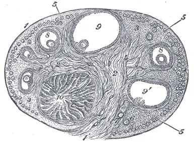
The ovary has an inner medullary core, and an outer cortical region, and is enclosed by a capsule of dense fibrous tissue the tunica albuginea. A layer of cuboidal epithelium from the visceral peritoneum covers the ovaries as ovarian surface epithelium.
The ovarian medulla is the highly vascular stromal tissue in the center of the ovary. Its contents include blood vessels, lymphatic vessels, immune cells, and nerves.
The ovarian hilus is the hilum in the ovary, formed by the stroma and attached to the ovarian ligament where the blood vessels, lymphatic vessels and nerves enter the medulla of the ovary.
It is also the main site of ovarian hilus cells also known as ovarian Leydig cells.
The ovarian cortex is an ill-defined outer region containing many ovarian follicles at different stages of development. The follicles are surrounded by peripheral stroma. It can be hard to distinguish between the cortex and medulla, but follicles are usually not found in the medulla.
Follicles
During prenatal development in the process of oogenesis – the development of the egg cell, follicles form consisting of primary oocytes surrounded by a layer of follicular cells. The earliest follicles are the primordial follicles that consist of a single layer of pre-granulosa cells known as follicular cells surrounding a primary oocyte. The follicles are surrounded by peripheral stromal tissue. A large number of primordial follicles are present at birth of which many will be lost perinatally. Follicles develop to maturity in a process of folliculogenesis that supports the development of the oocyte to the mature egg cell.
At puberty, in the menstrual cycle the primordial follicles become triggered to change to primary follicles, and the follicular cells change to granulosa cells. In the transition from primary follicle to secondary follicle, layers of granulosa cells, and an external covering known as the theca forms.
In the later stages of folliculogenesis the follicles include granulosa cells of the cumulus oophorus, and its inner layer of corona radiata, the zona pellucida, and the internal and external cell layers of the theca, and follicular fluid. The corpus luteum is the remains of the ruptured follicle after ovulation.
Blood supply
The ovarian artery arises from the abdominal aorta and supplies oxygenated blood to the ovary. The ovarian vein drains deoxygenated blood from the ovary to the inferior vena cava or one of its tributaries. The vessels are located within the suspensory ligament of the ovary.
Development
The ovaries develop in early embryogenesis from the genital ridge, following sexual differentiation. The sex of an organism is determined at the time of fertilization, but differentiation only takes place around six weeks later.
Function
The ovaries are the site of oogenesis and function to produce, develop and release the oocytes at ovulation for possible fertilisation. The ovary also produces hormones to assist in this process. The oocytes develop and mature to secondary oocytes in ovarian follicles. Each primordial follicle contains an oocyte surrounded by a single layer of pre-granulosa cells. At puberty, when the primordial follicle changes to a primary follicle, other layers of granulosa cells surround the follicle and become enclosed in a connective tissue covering called the theca. The follicles typically develop one at a time. As they develop they fill with follicular fluid which marks the secondary oocyte stage. When the fluid fills the antrum they are known as antral follicles, or pre-ovulatory follicles.
At puberty, the ovary begins to secrete increasing levels of hormones. Secondary sex characteristics begin to develop in response to the hormones. The ovary changes structure and function beginning at puberty. Since the ovaries can regulate hormones, they also play an important role in pregnancy and fertility. Usually, ovulation occurs in one of the two ovaries, releasing an egg cell each menstrual cycle. When an egg cell (secondary oocyte) is released and swept into the fallopian tube, feedback mechanisms stimulate the endocrine system, which causes hormone levels to change. These feedback mechanisms are controlled by the hypothalamus and pituitary glands. Messages or signals from the hypothalamus are sent to the pituitary gland. In turn, the pituitary gland releases hormones to the ovaries. From this signaling, the ovaries release their own hormones.
On maturing, a surge of luteinizing hormone is secreted by the pituitary gland, which stimulates the release of the oocyte through the rupture of the follicle, a process called ovulation. The remaining granulosa cells and theca cells from the ruptured follicle reorganize into the corpus luteum, which secretes progesterone to prepare the uterus for an eventual implantation of the embryo.
At maturity, ovaries secrete estrogen, androgen, inhibin, and progestogen. In women before menopause, 50% of testosterone is produced by the ovaries and released directly into the blood stream. The other 50% of testosterone in the bloodstream is made from the conversion of the adrenal pre-androgens (DHEA and androstenedione) to testosterone in other parts of the body. Estrogen is responsible for the appearance of secondary sex characteristics for females at puberty and for the maturation and maintenance of the reproductive organs in their mature functional state. Progesterone prepares the uterus for pregnancy and the mammary glands for lactation. Progesterone functions with estrogen by promoting menstrual cycle changes in the endometrium.
Clinical significance
Ovarian diseases can be classified as endocrine disorders or as a disorders of the reproductive system. Ovarian diseases include ovarian tumors such as ovarian germ cell tumors, ovarian cancers, and different types of ovarian cyst. Other ovarian disorders include inflammation (oophoritis), diminished functional activity (hypogonadism), failure to ovulate (annovulation), and twisting (ovarian torsion), or rupture (apoplexy) of the ovary. The ovaries may be affected by the growth of endometrial-like tissue on them (endometriosis).
If the egg fails to release from the follicle in the ovary, an ovarian cyst may form. Small ovarian cysts are common in healthy women. Some women have more follicles than usual caused by a hormonal disorder (polyendocrine metabolic ovarian syndrome), which inhibits the follicles from growing normally, and this will cause cycle irregularities.
The surgical removal or one or both ovaries is called an oophorectomy.
Some types of cancer therapy can negatively impact fertility. An option to preserve this is ovarian tissue cryopreservation where some of the tissue of the ovary cortex is removed and frozen to be replaced near to the remaining tissue after therapy completion. Tissue cryopreservation is also carried out in those who wish to postpone pregnancy, or to delay menopause. Another option to safeguard the ovaries during treatment is ovarian transposition which repositions the ovaries to a safer more shielded nearby location. Both options can be performed by laparoscopy.
Ovotesticular syndrome is a rare congenital condition of atypical sexual differentiation where an individual is born with both ovarian and testicular tissue. It is one of the rarest disorders of sex development, with an incidence rate of approximately one in 100,000 live births. Commonly, one or both gonads is an ovotestis containing both types of tissue. Although it is similar in some ways to mixed gonadal dysgenesis, the conditions can be distinguished histologically.
Source: Wikipedia, The Free Encyclopedia · CC BY-SA 4.0
References
- Ovary — Wikipedia, The Free EncyclopediaText from the Wikipedia article "Ovary" (CC BY-SA 4.0); excerpted. Licence. Retrieved 2026-10-19. Revision used.
Text above is quoted from the listed sources without changes to its meaning. Image credits and licences are shown with each image. Educational information only, not medical advice.
Related conditions
Educational anatomy reference, not medical advice. About this content.