
Pineal gland
Glandula pinealis
View the pineal gland in 3DThe pineal gland (also called the pineal body) is a small endocrine gland in the brain of most vertebrates. It produces melatonin, a serotonin-derived hormone, which modulates sleep patterns following the diurnal cycles. The shape of the gland resembles a pine cone, which gives it its name.
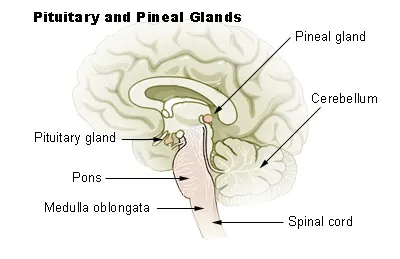
The pineal gland is located in the epithalamus, near the center of the brain, between the two hemispheres, tucked in a groove where the two halves of the thalamus join. It is one of the neuroendocrine secretory circumventricular organs in which capillaries are mostly permeable to solutes in the blood.
The pineal gland is present in almost all vertebrates, but is absent in protochordates, in which there is a simple pineal homologue. The hagfish, archaic vertebrates, lack a pineal gland. In some species of amphibians and reptiles, the gland is linked to a light-sensing organ, variously called the parietal eye, the pineal eye or the third eye. Reconstruction of the biological evolution pattern suggests that the pineal gland was originally a kind of atrophied photoreceptor that developed into a neuroendocrine organ.
Galen in the 2nd century C.E. could not find any functional role and regarded the gland as a structural support for the brain tissue. He gave the name konarion, meaning small cone or pinecone, which during the Renaissance was translated into Latin as pinealis. The 17th-century philosopher René Descartes regarded the gland as the primary locus of mind-body interaction, describing it as the "principal seat of the soul".
Structure
The pineal gland is a pine cone-shaped (hence the name), unpaired midline brain structure. It is reddish-gray in colour and about the size of a grain of rice (5–8 mm) in humans. It forms part of the epithalamus. It is attached to the rest of the brain by a pineal stalk. The ventral lamina of the pineal stalk is continuous with the posterior commissure, and its dorsal lamina with the habenular commissure.
Location
It normally lies in a depression between the two superior colliculi. It is situated between the laterally positioned thalamic bodies, and posterior to the habenular commissure. It is located in the quadrigeminal cistern behind the third ventricle, enclosing the pineal recess.
Blood supply
Unlike most of the mammalian brain, the pineal gland is not isolated from the body by the blood–brain barrier system; it has profuse blood flow, second only to the kidney, supplied from the choroidal branches of the posterior cerebral artery.
Afferents
The afferent nerve supply of pineal gland is by the nervus conarii which receives postganglionic sympathetic afferents from the superior cervical ganglion, and parasympathetic afferents from the pterygopalatine ganglia and otic ganglia. According to research on animals, neurons of the trigeminal ganglion that are involved in pituitary adenylate cyclase-activating peptide neuropeptide signaling project the gland.
Neural pathway for melatonin production
The canonical neural pathway regulating pineal melatonin production begins in the eye with the intrinsically photosensitive ganglion cells of the retina which project inhibitory GABAergic efferents to the paraventricular nucleus of hypothalamus via the retinohypothalamic tract. The paraventricular nucleus in turn projects to the superior cervical ganglia, which finally projects to the pineal gland. Darkness thus leads to disinhibition of the paraventricular nucleus, leading it to activate pineal gland melatonin production by way of the superior cervical ganglia.
Microanatomy
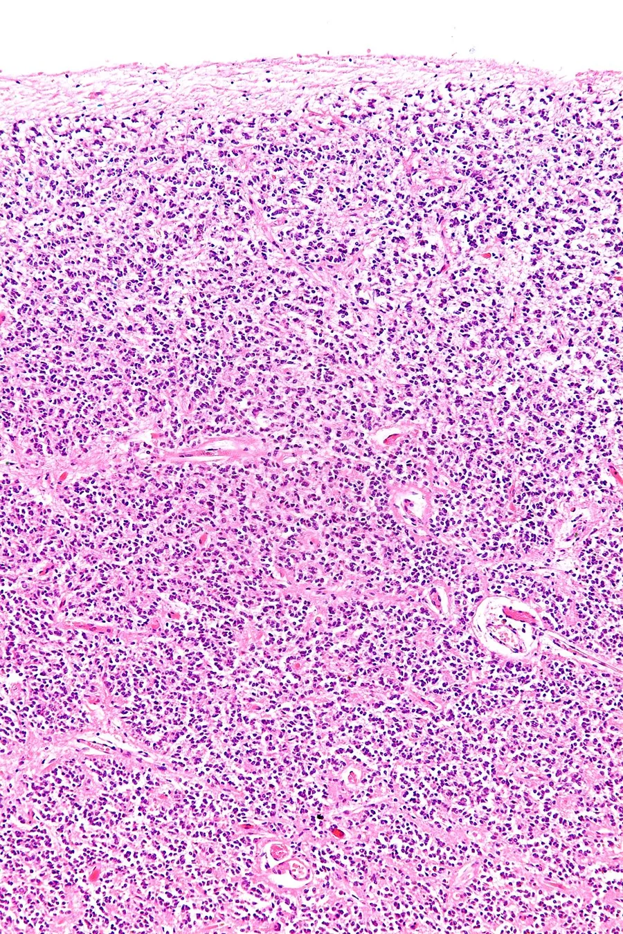
The pineal body in humans consists of a lobular parenchyma of pinealocytes surrounded by connective tissue spaces. The gland's surface is covered by a pial capsule.
The pineal gland consists mainly of pinealocytes, but four other cell types have been identified. As it is quite cellular (in relation to the cortex and white matter), it may be mistaken for a neoplasm.
Development
The human pineal gland grows in size until about 1–2 years of age, remaining stable thereafter, although its weight increases gradually from puberty onwards. The abundant melatonin levels in children are believed to inhibit sexual development, and pineal tumors have been linked with precocious puberty. When puberty arrives, melatonin production is reduced.
Symmetry
In the zebrafish the pineal gland does not straddle the midline, but shows a left-sided bias. In humans, functional cerebral dominance is accompanied by subtle anatomical asymmetry.
Function
A principal function of the pineal gland is to produce melatonin, which has various functions in the central nervous system, the most important of which is to help modulate sleep patterns; melatonin production is stimulated by darkness and inhibited by light. Intrinsically photosensitive retinal ganglion cells in the retina detect light and send this signal to the suprachiasmatic nucleus (SCN), synchronizing the SCN to the day-night cycle. Fibers then relay the daylight information to the paraventricular nuclei and spinal cord via the sympathetic nervous system to the superior cervical ganglia, and then to the pineal gland.
Regulation of the pituitary gland
Studies on rodents suggest that the pineal gland influences the pituitary gland's secretion of the sex hormones, follicle-stimulating hormone (FSH), and luteinizing hormone (LH). Pinealectomy performed on rodents produced no change in pituitary weight, but caused an increase in the concentration of FSH and LH within the gland.
The pineal gland contains receptors for the regulatory neuropeptide endothelin 1, which, when injected in picomolar quantities into the lateral cerebral ventricle, causes a calcium-mediated increase in pineal glucose metabolism.
Calcification
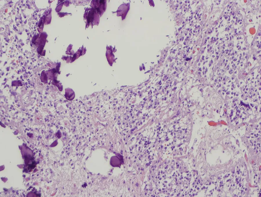
Calcification of the pineal gland is typical in young adults, and has been observed in children as young as two years of age. Pineal gland calcification is detrimental to its ability to synthesize melatonin, although it remains inconclusive whether pineal calcification causes sleep problems.
The calcified gland is often seen in skull X-rays. Calcification rates vary widely by country and correlate with an increase in age, with calcification occurring in an estimated 40% of Americans by age seventeen. Calcification of the pineal gland is associated with corpora arenacea, also known as "brain sand".
Tumors
Tumors of the pineal gland are called pinealomas. These tumors are rare and 50% to 70% are germinomas that arise from sequestered embryonic germ cells. Histologically they are similar to testicular seminomas and ovarian dysgerminomas.
A pineal tumor can compress the superior colliculi and pretectal area of the dorsal midbrain, producing Parinaud's syndrome. Pineal tumors also can cause compression of the cerebral aqueduct, resulting in a noncommunicating hydrocephalus. Other manifestations are the consequence of their pressure effects and consist of visual disturbances, headache, mental deterioration, and sometimes dementia-like behaviour.
These neoplasms are divided into three categories: pineoblastomas, pineocytomas, and mixed tumors, based on their level of differentiation, which, in turn, correlates with their neoplastic aggressiveness. The clinical course of patients with pineocytomas is prolonged, averaging up to several years. The position of these tumors makes them difficult to remove surgically.
Other conditions
The morphology of the pineal gland differs markedly in different pathological conditions. For instance, it is known that its volume is reduced in both obese patients and those with primary insomnia.
Source: Wikipedia, The Free Encyclopedia · CC BY-SA 4.0
References
- Pineal gland — Wikipedia, The Free EncyclopediaText from the Wikipedia article "Pineal gland" (CC BY-SA 4.0); excerpted. Licence. Retrieved 2026-10-19. Revision used.
Text above is quoted from the listed sources without changes to its meaning. Image credits and licences are shown with each image. Educational information only, not medical advice.
Educational anatomy reference, not medical advice. About this content.