
Sphenoid bone
Os sphenoidale
View the sphenoid bone in 3DThe sphenoid bone is an unpaired bone of the neurocranium in mammals. It is situated in the middle of the skull towards the front, in front of the basilar part of the occipital bone. The sphenoid bone is one of the seven bones that articulate to form the orbit. The name originates from the Ancient Greek word sphekodes (σφηκώδης), meaning 'wasp-like', presumably referring to the bone's winged appearance.
Structure
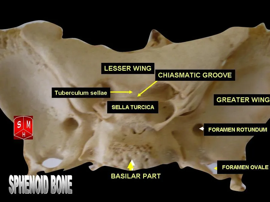
a median portion, known as the body of sphenoid bone, containing the sella turcica, which houses the pituitary gland as well as the paired paranasal sinuses, the sphenoidal sinuses
two greater wings on the lateral side of the body and two lesser wings from the anterior side.
Pterygoid processes of the sphenoid, directed downwards from the junction of the body and the greater wings.
Two sphenoidal conchae are situated at the anterior and inferior part of the body.
Intrinsic ligaments of the sphenoid
the pterygospinous, which stretches between the spina angularis and the lateral pterygoid plate, superiorly attached to the root of the lateral pterygoid plate anteriorly;
the interclinoid ligament, a band of dura mater that connects the anterior clinoid process and posterior clinoid process of the sphenoid bone;
the caroticoclinoid, connecting the anterior to the middle clinoid process.
These ligaments occasionally ossify, though the incidence of ligamentous ossification (both partial and complete) varies according to the ligament type, with the interclinoid ligament being most commonly identified as having ossified and the pterygoalar ligament least commonly identified.
Articulations
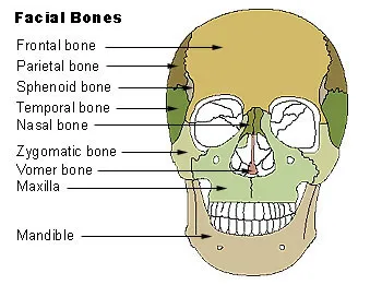
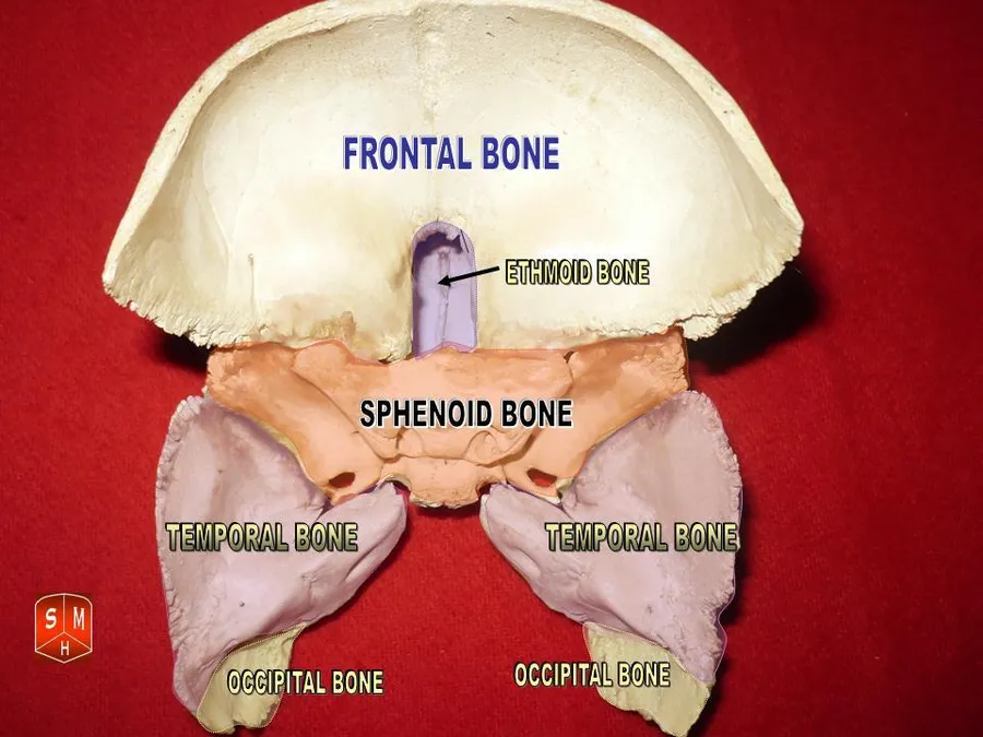
The sphenoid articulates with the frontal, parietal, ethmoid, temporal, zygomatic, palatine, vomer, and occipital bones and helps to connect the neurocranium to the facial skeleton.
Function
This bone assists with the formation of the base and the sides of the skull and the floors and walls of the orbits. It is the site of attachment for most of the muscles of mastication. Many foramina and fissures are located in the sphenoid that carry nerves and blood vessels of the head and neck, such as the superior orbital fissure (with ophthalmic nerve), foramen rotundum (with maxillary nerve) and foramen ovale (with mandibular nerve).
Source: Wikipedia, The Free Encyclopedia · CC BY-SA 4.0
References
- Sphenoid bone — Wikipedia, The Free EncyclopediaText from the Wikipedia article "Sphenoid bone" (CC BY-SA 4.0); excerpted. Licence. Retrieved 2026-10-19. Revision used.
Text above is quoted from the listed sources without changes to its meaning. Image credits and licences are shown with each image. Educational information only, not medical advice.
Educational anatomy reference, not medical advice. About this content.