Age-related macular degeneration
Also called: AMD, Wet AMD, Dry AMD
Macular degeneration, or age-related macular degeneration (AMD), is a leading cause of vision loss in Americans 60 and older. It is a disease that destroys your sharp, central vision. You need central vision to see objects clearly and to do tasks such as reading and driving.
AMD affects the macula, the part of the eye that allows you to see fine detail. It does not hurt, but it causes cells in the macula to die. There are two types: wet and dry. Wet AMD happens when abnormal blood vessels grow under the macula. These new blood vessels often leak blood and fluid. Wet AMD damages the macula quickly. Blurred vision is a common early symptom. Dry AMD happens when the light-sensitive cells in the macula slowly break down. You gradually lose your central vision. A common early symptom is that straight lines appear crooked.
Regular comprehensive eye exams can detect macular degeneration before the disease causes vision loss. Treatment can slow vision loss. It does not restore vision.
NIH: National Eye Institute

Signs and symptoms
Early or intermediate AMD may be asymptomatic, or it may present with blurred or decreased vision in one or both eyes. This may manifest initially as difficulty with reading or driving (especially in poorly lit areas). Other symptoms of AMD include distortion of vision and blind spots (especially in and around the central visual field).
Other signs and symptoms of macular degeneration include:
Distorted vision in the form of metamorphopsia, in which a grid of straight lines appears wavy and parts of the grid may appear blank: Patients often first notice this when looking at things like miniblinds in their home or telephone poles while driving.
Central scotomas, shadows, or missing areas of vision
Slow recovery of visual function after exposure to bright light (photostress test)
Visual acuity drastically decreasing. (two levels or more), e.g.: 20/20 to 20/80
Blurred vision: Those with nonexudative (dry) macular degeneration may be asymptomatic or notice a gradual loss of central vision, whereas those with exudative (wet) macular degeneration often notice a rapid onset of vision loss (often caused by leakage and bleeding of abnormal blood vessels).
Trouble discerning colors, specifically dark ones from dark ones and light ones from light ones
Formed visual hallucinations and flashing lights have also been associated with severe visual loss secondary to wet AMD.
Macular degeneration by itself will not lead to total blindness. For that matter, only a small number of people with visual impairment are blind. In almost all cases, some vision remains, mainly peripheral. Other complicating conditions may lead to such an acute condition (severe stroke or trauma, untreated glaucoma, etc.), but few macular degeneration patients experience total visual loss.
The area of the macula constitutes only about 2.1% of the retina, and the remaining 97.9% (the peripheral field) remains unaffected by the disease. Even though the macula provides such a small fraction of the visual field, almost half of the visual cortex is devoted to processing macular information.
In addition, people with dry macular degeneration often do not experience any symptoms but can experience gradual onset of blurry vision in one or both eyes. People with wet macular degeneration may experience acute onset of visual symptoms.
Risk factors
The pathogenesis of age-related macular degeneration (AMD) is intricately linked to gene-environment interactions. Key risk factors are age, race/ethnicity, smoking, and family history. Advanced age is the strongest predictor of AMD, particularly over 50.
Race and ethnicity
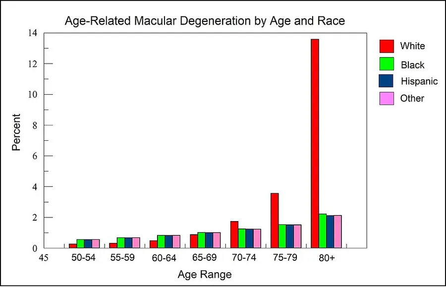
As illustrated by the Figure in this section, derived from data presented by the National Eye Institute of the United States, among those over 80 years of age, white individuals are more than 6-fold more likely to develop AMD than Black or Hispanic individuals. Thus, white background is a major risk factor for AMD.
In Caucasian (white) skin, there is a specific group of polymorphic genes (with single nucleotide alterations) that encode for enzymes and transcription factors responsible for the early steps (including the first step, formation of L-DOPA from the amino acid tyrosine) of the melanin synthesis pathway. Many of these enzymes and transcription factors are reviewed by Markiewicz and Idowu. Also, as reviewed by Sturm et al. "increasing intracellular concentrations of either tyrosine or L-DOPA both increase melanogenesis" or formation of the black pigment melanin. Thus there appears to be an association between reduced L-DOPA production and white skin. As suggested by the Figure and information in this section, reduced L-DOPA, resulting in white skin, appears to be associated with an increased risk of macular degeneration for white individuals over the age of 80.
Environment and lifestyle
Smoking: Smoking tobacco increases the risk of AMD by two to three times that of someone who has never smoked, and may be the most important modifiable factor in its prevention. A review of previous studies found "a strong association between current smoking and AMD. ... Cigarette smoking is likely to have toxic effects on the retina."
Hypertension (high blood pressure): In the ALIENOR study 2013, early and late AMD were not significantly associated with systolic or diastolic blood pressure (BP), hypertension, or use of antihypertensive medications, but elevated pulse pressure [(PP) systolic BP minus diastolic BP] was significantly associated with an increased risk of late AMD.
High cholesterol: Elevated cholesterol may increase the risk of AMD
Obesity: Abdominal obesity is a risk factor, especially among men
Fat intake: Consuming high amounts of certain fats, including saturated fats, trans fats, and omega-6 fatty acids, likely contributes to AMD, while monounsaturated fats are potentially protective. In particular, omega-3 fatty acids may decrease the risk of AMD.
Exposure to UV light from sunlight may be associated with an increased risk of developing AMD, although evidence for this is weaker than for other causes.
There is no evidence to support the claim that exposure to digital screens contributes to the risk of macular degeneration.
Genetics
AMD is a highly heritable condition. Recurrence ratios for siblings of an affected individual are three- to six-fold higher than in the general population. Genetic linkage analysis has identified 5 sets of gene variants at three locations on different chromosomes (1, 6 and 10) as explaining at least 50% of the risk. These genes have roles regulating the immune response, inflammatory processes and homeostasis of the retina. Variants of these genes give rise to different kinds of dysfunction in these processes. Over time, this results in the accumulation of intracellular and extracellular metabolic debris. This can cause scarring of the retina or breakdown of its vascularization.
The list of genetic variations associated with AMD includes complement factors, apolipoprotein E, fibroblast growth factor 2, DNA excision repair protein, and age-related maculopathy susceptibility protein 2.
Although genetic testing can lead to the identification of genetic variation that can predispose to AMD, the complex pathogenesis of the condition prevents the use of these tests in routine practice. Nevertheless, they can be useful in selecting patients for clinical trials and analyzing their response to treatment. The three loci where identified gene variants are found are designated:
Complement factor H (CFH) on chromosome 1 at location 1q31.3
HTRA serine peptidase 1/Age Related Maculopathy Susceptibility 2 (HTRA1/ARMS2) on chromosome 10 at location 10q26
Complement factor B/complement component 2 (CFB/CC2) on chromosome 6 at 6p21.3
Specific genes
Polymorphisms in genes for complement system proteins: Variation in the genes for the complement system proteins factor H (CFH), factor B (CFB) and factor 3 (C3), among others, are strongly associated with a person's risk for developing AMD. CFH is involved in inhibiting the inflammatory response. The mutation in CFH (Y402H) results in reduced ability of the protein to localise to and protect tissues such as the retina from complement overactivation. Absence of the complement factor H-related genes R3 and R1 protects against AMD. Two independent studies in 2007 showed a certain common mutation Arg80Gly in the C3 gene, which is a central protein of the complement system, is strongly associated with the occurrence of AMD. The authors of both papers consider their study to underscore the influence of the complement pathway in the pathogenesis of this disease.
In two 2006 studies, another gene that has implications for the disease, called HTRA1 (encoding a secreted serine protease), was identified.
Six mutations of the gene SERPING1 (Serpin Peptidase Inhibitor, Clade G (C1 Inhibitor), Member 1) are associated with AMD. Mutations in this gene can also cause hereditary angioedema.
Fibulin-5 mutation: Rare forms of the disease are caused by genetic defects in fibulin-5, in an autosomal dominant manner. In 2004, Stone et al. performed a screening on 402 AMD patients and revealed a statistically significant correlation between mutations in fibulin-5 and the incidence of the disease.
Mitochondrial-related gene polymorphisms such as that in the MT-ND2 molecule, predicts wet AMD.
Diagnosis
Diagnosis of age-related macular degeneration depends on signs in the macula, not necessarily vision. Early diagnosis of AMD can prevent further visual deterioration and potentially improve vision.
Diagnosis of dry (or early stage) AMD may include the following clinical examinations as well as procedures and tests:
The transition from dry to wet AMD can happen rapidly, and if it is left untreated can lead to legal blindness in as little as six months. To prevent this from occurring and to initiate preventive strategies earlier in the disease process, dark adaptation testing may be performed. A dark adaptometer can detect subclinical AMD at least three years earlier than it is clinically evident.
There is a loss of contrast sensitivity, so that contours, shadows, and color vision are less vivid. The loss of contrast sensitivity can be quickly and easily measured by a contrast sensitivity test like Pelli Robson performed either at home or by an eye specialist.
When viewing an Amsler grid, some straight lines appear wavy and some patches appear blank
When viewing a Snellen chart, at least 2 lines decline
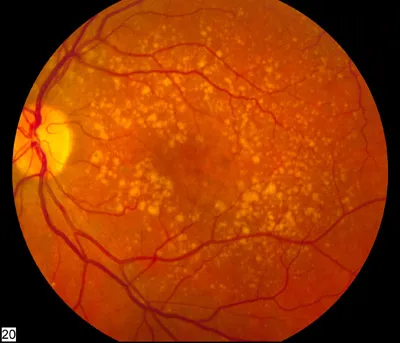
In dry macular degeneration, which occurs in 85–90 percent of AMD cases, drusen spots can be seen in Fundus photography
Using an electroretinogram, points in the macula with a weak or absent response compared to a normal eye may be found
Farnsworth-Munsell 100 hue test and Maximum Color Contrast Sensitivity test (MCCS) for assessing color acuity and color contrast sensitivity
Optical coherence tomography is now used by most ophthalmologists in the diagnosis and the follow-up evaluation of the response to treatment with antiangiogenic drugs.
Diagnosis of wet (or late-stage) AMD may include the following in addition to the above tests:
Preferential hyperacuity perimetry changes (for wet AMD). Preferential hyperacuity perimetry is a test that detects drastic changes in vision and involves the macula being stimulated with distorted patterns of dots and the patient identification of where in the visual field this occurs.
In wet macular degeneration, angiography can visualize the leakage of the bloodstream behind the macula. Fluorescein angiography allows for the identification and localization of abnormal vascular processes.
Histology
Pigmentary changes in the retina – In addition to the pigmented cells in the iris (the colored part of the eye), there are pigmented cells beneath the retina. As these cells break down and release their pigment, dark clumps of released pigment and later, areas that are less pigmented may appear
Exudative changes: hemorrhages in the eye, hard exudates, subretinal/sub-RPE/intraretinal fluid
Drusen, tiny accumulations of extracellular material that build up on the retina. While there is a tendency for drusen to be blamed for the progressive loss of vision, drusen deposits can be present in the retina without vision loss. Some patients with large deposits of drusen have normal visual acuity. If normal retinal reception and image transmission are sometimes possible in a retina when high concentrations of drusen are present, then, even if drusen can be implicated in the loss of visual function, there must be at least one other factor that accounts for the loss of vision.
Management
Treatment of AMD varies depending on the category of the disease at the time of diagnosis. In general, treatment is aimed at slowing down the progression of AMD. As of 2018, there are no treatments to reverse the effects of AMD. As of 2024, there are two drugs to dissolve the drusen in dry AMD, see below. Early-stage and intermediate-stage AMD is managed by modifying known risk factors such as smoking cessation, management of hypertension and atherosclerosis, and making dietary modifications. For intermediate-stage AMD, management also includes antioxidant and mineral supplementation. Advanced-stage AMD is managed based on the presence of choroidal neovascularization (CNV): dry AMD (no CNV present) or wet AMD (CNV present). No effective treatments exist for dry AMD. The CNV present in wet AMD is managed with vascular endothelial growth factor (VEGF) inhibitors. Daily use of an Amsler grid or other home visual monitoring tools can be used to monitor for development of distorted vision, which may be a sign of disease progression.
Dietary supplements
Dietary supplements may be suggested for people with AMD, to reduce damage to the cells in the retina with antioxidants. The formulations commonly suggested are known as AREDS. The specific vitamins and minerals in AREDS-1 are vitamin C (500 mg), zinc (80 mg), vitamin E (400 IU), copper (2 mg) and beta-carotene (15 mg). In the AREDS-2 formulation, lutein (10 mg) and zeaxanthin (2 mg) replaced beta-carotene due to the risk of lung cancer in smokers taking beta-carotene. There is some evidence to indicate that people with bilateral early or intermediate AMD, or intermediate AMD in one eye and advanced AMD in the other eye may benefit from vitamin and mineral supplementation. AREDS supplementation may help slow the progression to more severe forms of AMD and there is some evidence of improved visual acuity at 5 years. There is no evidence that micronutrient supplementation prevents AMD progression in those with severe disease or prevents disease onset in those without AMD.
With regards to AREDS-1 compared with AREDS-2 formulations, there is only weak evidence comparing the effectiveness of each formulation and the effectiveness of lutein and zeaxanthin as a replacement in the AREDS-2 formulation.
Dry AMD
Pegcetacoplan (Syfovre) and avacincaptad pegol (Izervay) are approved for medical use in the United States. In 2023 it was reported that the aging pigment lipofuscin can be broken down with the help of melanin and drugs through a newly discovered mechanism. The pigment lipofuscin plays a central role in the development of dry AMD and Stargardt's disease. The clinical development of this mechanism, which has the potential to clear Bruch's membrane and reduce the formation of drusen, is in preparation.
Wet AMD
Bevacizumab, ranibizumab, aflibercept, brolucizumab, and faricimab are approved VEGF inhibitors for the treatment of CNV in wet AMD. All these drugs are administered via intravitreal injection, meaning they are injected directly into the eye. Bevacizumab is another VEGF inhibitor that has been shown to have similar efficacy and safety. FDA approved ophthalmic bevacizumab for wet AMD 7/24/2026 Outlook Therapeutics’ Lytenava.AMD can also be treated with laser coagulation therapy.
The American Academy of Ophthalmology practice guidelines do not recommend laser coagulation therapy for macular degeneration, but state that it may be useful in people with new blood vessels in the choroid outside of the fovea who do not respond to drug treatment. There is strong evidence that laser coagulation will result in the disappearance of drusen but does not affect choroidal neovascularisation. A 2007 Cochrane review found that laser photocoagulation of new blood vessels in the choroid outside of the fovea is an effective and economical method, but that the benefits are limited for vessels next to or below the fovea.
Photodynamic therapy has also been used to treat wet AMD. The drug verteporfin is administered intravenously; light of a certain wavelength is then applied to the abnormal blood vessels. This activates the verteporfin destroying the vessels.
Cataract surgery could improve visual outcomes for people with AMD, though there have been concerns about surgery increasing the progression of AMD. A randomized controlled trial found that people who underwent immediate cataract surgery (within two weeks) had improved visual acuity and better quality of life outcomes than those who underwent delayed cataract surgery (6 months).
Radiotherapy has been proposed as a treatment for wet AMD but the evidence to support the use of modern stereotactic radiotherapy combined with anti-VEGF is currently uncertain and is awaiting the results of ongoing studies.
Adaptive devices
Because peripheral vision is not affected, persons with macular degeneration can learn to use their remaining vision to partially compensate. Assistance and resources are available in many countries and every state in the U.S. Classes for "independent living" are given and some technology can be obtained from a state department of rehabilitation.
Adaptive devices can help people read. These include magnifying glasses, special eyeglass lenses, computer screen readers, electronic glasses, and TV systems that enlarge the reading material.
Computer screen readers such as JAWS or Thunder work with standard Windows computers.
Also, Apple devices provide a wide range of features (voice-over, screen readers, Braille, etc.).
Video cameras can be fed into standard or special-purpose computer monitors, and the image can be zoomed in and magnified. These systems often include a movable table to move the written material.
Accessible publishing provides larger fonts for printed books, patterns to make tracking easier, audiobooks and DAISY books with both text and audio.
Epidemiology
The prevalence of any age-related macular degeneration is higher in Europeans than in Asians and Africans. There is no difference in prevalence between Asians and Africans. The incidence of age-related macular degeneration and its associated features increases with age and is low in people <55 years of age. Smoking is the strongest modifiable risk factor. As of 2008, age-related macular degeneration accounts for more than 54% of all vision loss in the white population in the US. An estimated 8 million Americans are affected with early age-related macular degeneration, of whom over 1 million will develop advanced age-related macular degeneration within the next 5 years. In the UK, age-related macular degeneration is the cause of blindness in almost 42% of those who go blind aged 65–74 years, almost two-thirds of those aged 75–84 years, and almost three-quarters of those aged 85 years or older.
Source: Wikipedia, The Free Encyclopedia · CC BY-SA 4.0
References
- Macular Degeneration — MedlinePlus, U.S. National Library of MedicineSource: MedlinePlus, National Library of Medicine Public domain (U.S. government work). Retrieved 2026-10-19.
- Macular degeneration — Wikipedia, The Free EncyclopediaText from the Wikipedia article "Macular degeneration" (CC BY-SA 4.0); excerpted. Licence. Retrieved 2026-10-19. Revision used.
Text above is quoted from the listed sources without changes to its meaning. Image credits and licences are shown with each image. Educational information only, not medical advice.
Related conditions
Educational overview, not medical advice. Treatment varies by person and location. Consult a qualified health professional for diagnosis or care. Content and model notes · Privacy information