
Navicular
Os naviculare sinistrum
- Paired structure: one on the left and one on the right side of the body.
The navicular bone is a small bone found in the feet of most mammals.
Human anatomy
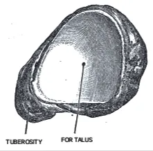
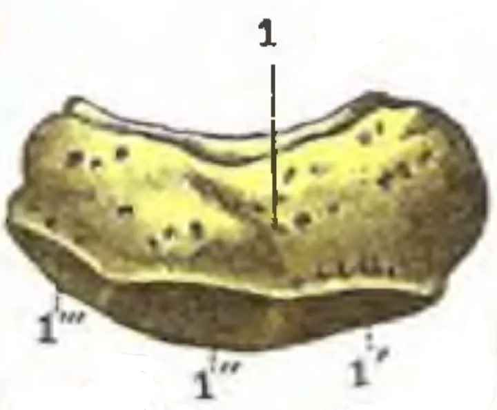
The navicular bone in humans is one of the tarsal bones, found in the foot. Its name derives from the human bone's resemblance to a small boat, caused by the strongly concave proximal (i.e., posterior) articular surface. The term navicular bone or hand navicular bone was historically used for the scaphoid bone, one of the carpal bones of the wrist.
The navicular bone in humans is located on the medial side of the foot, and articulates proximally with the talus, distally with the three cuneiform bones, and laterally with the cuboid.
It is the last of the foot bones to start ossification and does not tend to do so until the beginning of the fourth year in typical anatomy, although a large range of variation has been reported.
The tibialis posterior is the only muscle that attaches to the navicular bone. The main portion of the muscle inserts into the tuberosity of the navicular bone. An accessory navicular bone may be present in 2–14% of the general population.
Clinical significance
The human navicular is not a commonly broken bone but it breaks due to two reasons. The first mechanism is a stress fracture which happens commonly in athletes, and the other mechanism is a high energy trauma.
The navicular bone is a keystone of the foot: it is part of the coxa pedis and articulates with the talus, first, second and third cuneiform, cuboid and calcaneus. It plays an important role in the biomechanics of the foot, helping in inversion, eversion, and motion; it is a structural link between midfoot and forefoot and it is part of the longitudinal and transverse arch of the foot.
Source: Wikipedia, The Free Encyclopedia · CC BY-SA 4.0
References
- Navicular bone — Wikipedia, The Free EncyclopediaText from the Wikipedia article "Navicular bone" (CC BY-SA 4.0); excerpted. Licence. Retrieved 2026-10-19. Revision used.
Text above is quoted from the listed sources without changes to its meaning. Image credits and licences are shown with each image. Educational information only, not medical advice.
Educational anatomy reference, not medical advice. About this content.