
Palatine bone
Os palatinum sinistrum
- Paired structure: one on the left and one on the right side of the body.
In anatomy, the palatine bones (derived from the Latin palatum) are two irregular bones of the facial skeleton in many animal species, located above the uvula in the throat. Together with the maxilla, they comprise the hard palate.
Among fishes, it is often known as the dermopalatine to distinguish it from the autopalatine.
Structure
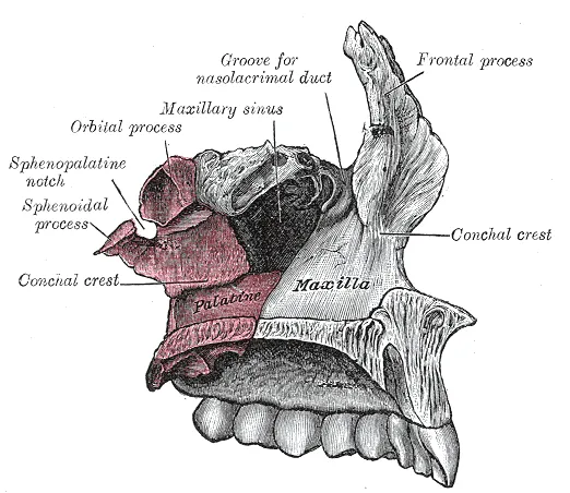
The palatine bones are situated at the back of the nasal cavity between the maxilla and the pterygoid process of the sphenoid bone.
They contribute to the walls of three cavities: the floor and lateral walls of the nasal cavity, the roof of the mouth, and the floor of the orbits. They help to form the pterygopalatine and pterygoid fossae, and the inferior orbital fissures.
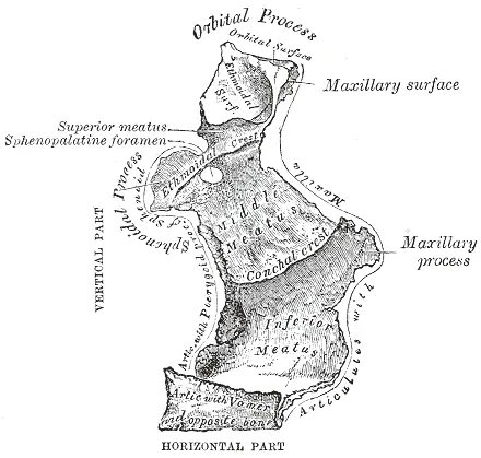
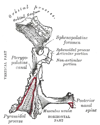
Each palatine bone somewhat resembles the letter L, and consists of a horizontal plate, a perpendicular plate, and three projecting processes—the pyramidal process, which is directed backward and lateral from the junction of the two parts, and the orbital and sphenoidal processes, which surmount the vertical part, and are separated by a deep notch, the sphenopalatine notch. The two plates form the posterior part of the hard palate and the floor of the nasal cavity; anteriorly, they join with the maxillae. The two horizontal plates articulate with each other at the posterior part of the median palatine suture and more anteriorly with the maxillae at the transverse palatine suture.
The human palatine articulates with six bones: the sphenoid, ethmoid, maxilla, inferior nasal concha, vomer and opposite palatine.
There are two important foramina in the palatine bones that transmit nerves and blood vessels to this region: the greater and lesser palatine. The larger greater palatine foramen is located in the posterolateral region of each of the palatine bones, usually at the apex of the maxillary third molar. The greater palatine foramen transmits the greater palatine nerve and blood vessels. A smaller opening nearby, the lesser palatine foramen, transmits the lesser palatine nerve and blood vessels to the soft palate and tonsils. Both foramina are openings of the pterygopalatine canal that carries the descending palatine nerves and blood vessels from the pterygopalatine fossa to the palate.
Function
The sphenopalatine foramen is the opening between the sphenoid bone and orbital processes of the palatine bone; it opens into the nasal cavity and gives passage to branches from the pterygopalatine ganglion and the sphenopalatine artery from the maxillary artery.
Source: Wikipedia, The Free Encyclopedia · CC BY-SA 4.0
References
- Palatine bone — Wikipedia, The Free EncyclopediaText from the Wikipedia article "Palatine bone" (CC BY-SA 4.0); excerpted. Licence. Retrieved 2026-10-19. Revision used.
Text above is quoted from the listed sources without changes to its meaning. Image credits and licences are shown with each image. Educational information only, not medical advice.
Educational anatomy reference, not medical advice. About this content.