
COVID-19
Also called: Coronavirus disease 2019, SARS-CoV-2
Coronavirus disease 2019 (COVID-19) is a contagious disease caused by the coronavirus SARS-CoV-2. Starting in January 2020, the disease spread worldwide, resulting in the COVID-19 pandemic. In March 2020, the World Health Organization declared COVID-19 a global health emergency; they declared the end of the emergency on 5 May 2023.
The symptoms of COVID‑19 can vary but often include fever, fatigue, cough, shortness of breath, loss of smell, and loss of taste. Symptoms may begin one to fourteen days after exposure to the virus. At least a third of people who are infected do not develop noticeable symptoms. Of those who develop symptoms noticeable enough to be classified as patients, most (81%) develop mild to moderate symptoms (up to mild pneumonia), while 14% develop severe symptoms (dyspnea, hypoxia, or more than 50% lung involvement on imaging), and 5% develop critical symptoms (respiratory failure, shock, or multiorgan dysfunction). Older people have a higher risk of developing severe symptoms and dying. Some people experience persistent symptoms (long COVID), for months or years after infection, including fatigue, cognitive issues and shortness of breath. Damage to organs has been observed in a subset.
COVID‑19 transmission occurs when infectious particles are breathed in or come into contact with the eyes, nose, or mouth. The risk is highest when people are close together, but small airborne particles containing the virus can remain suspended in the air and travel over longer distances, particularly indoors. Transmission can also occur when people touch their eyes, nose, or mouth after touching surfaces or objects that have been contaminated by the virus. People remain contagious for up to 20 days and can spread the virus even if they do not develop symptoms.
There are two common tests to detect a COVID infection. Antigen tests (also called rapid lateral flow tests) can be used at home. A positive test indicates an active infection. However, negative test results are not always accurate, especially when there are no symptoms. Health care providers can perform a more accurate PCR test, which is typically analysed in a laboratory.
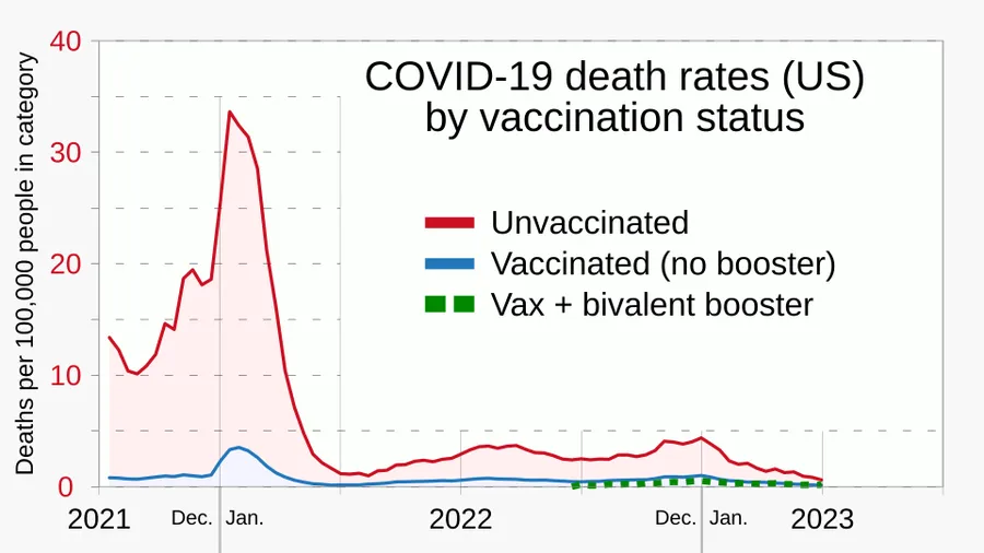
Several COVID-19 vaccines have been approved and distributed in various countries, many of which have initiated mass vaccination campaigns. Other preventive measures include physical or social distancing, quarantining, ventilation of indoor spaces, use of face masks or coverings in public, covering coughs and sneezes, hand washing, and keeping unwashed hands away from the face. Initial treatment consists of drugs that have been developed to inhibit the virus for those at high risk and symptomatic treatment, managing the disease through supportive care.
The first known case was identified in Wuhan, China, in December 2019. Most scientists believe that the SARS-CoV-2 virus entered into human populations through natural zoonosis, similar to the SARS-CoV-1 and MERS-CoV outbreaks, and consistent with other pandemics in human history. Social and environmental factors including climate change, natural ecosystem destruction and wildlife trade increased the likelihood of such zoonotic spillover.

Complications
Complications may include pneumonia, acute respiratory distress syndrome (ARDS), multi-organ failure, septic shock, and death. Cardiovascular complications may include heart failure, arrhythmias (including atrial fibrillation), heart inflammation, thrombosis, particularly venous thromboembolism, and endothelial cell injury and dysfunction. Approximately 20–30% of people who present with COVID‑19 have elevated liver enzymes, reflecting liver injury.
Neurological manifestations include seizure, stroke, encephalitis, and Guillain–Barré syndrome (which includes loss of motor functions). Following the infection, children may develop paediatric multisystem inflammatory syndrome, which has symptoms similar to Kawasaki disease, which can be fatal. In very rare cases, acute encephalopathy can occur, and it can be considered in those who have been diagnosed with COVID‑19 and have an altered mental status.
According to the US Centers for Disease Control and Prevention, pregnant women are at increased risk of becoming seriously ill from COVID‑19. This is because pregnant women with COVID‑19 appear to be more likely to develop respiratory and obstetric complications that can lead to miscarriage, premature delivery and intrauterine growth restriction.
Fungal infections such as aspergillosis, candidiasis, cryptococcosis and mucormycosis have been recorded in people recovering from COVID‑19.
Cause
COVID‑19 is caused by infection with a coronavirus known as "severe acute respiratory syndrome coronavirus 2" (SARS-CoV-2).
Virology
Severe acute respiratory syndrome coronavirus 2 (SARS-CoV-2) is a novel severe acute respiratory syndrome coronavirus. It was first isolated from three people with pneumonia connected to the cluster of acute respiratory illness cases in Wuhan. All structural features of the novel SARS-CoV-2 virus particle occur in related coronaviruses in nature, particularly in Rhinolophus sinicus (Chinese horseshoe bats).
Outside the human body, the virus is destroyed by household soap which bursts its protective bubble. Hospital disinfectants, alcohols, heat, povidone-iodine, and ultraviolet-C (UV-C) irradiation are also effective disinfection methods for surfaces.
SARS-CoV-2 is closely related to the original SARS-CoV. The virus is thought to have an animal (zoonotic) origin. Genetic analysis has revealed that the coronavirus genetically clusters with the genus Betacoronavirus, in subgenus Sarbecovirus (lineage B) together with two bat-derived strains. It is 96% identical at the whole genome level to other bat coronavirus samples (BatCov RaTG13). The structural proteins of SARS-CoV-2 include membrane glycoprotein (M), envelope protein (E), nucleocapsid protein (N), and the spike protein (S). The M protein of SARS-CoV-2 is about 98% similar to the M protein of bat SARS-CoV, maintains around 98% homology with pangolin SARS-CoV, and has 90% homology with the M protein of SARS-CoV; whereas, the similarity is only around 38% with the M protein of MERS-CoV.
SARS-CoV-2 variants
The SARS-CoV-2 virus that causes COVID-19 has continuously evolved since the start of the pandemic. Once a large number of mutations have accumulated, a subtype is classified as a variant. Major variants of concern were given a name based on letters of the Greek alphabet, for example, Alpha, Beta, Delta; after naming the Omicron variant in November 2021, the WHO stopped naming new variants of concern. The Pango system further specifies the lineage of the variant, for instance, JN.1 for a variant active in 2024, and XFG for one in 2025.
The Alpha variant (B.1.1.7) first appeared around autumn 2020. By the end of 2020, two other variants of concern had appeared: Gamma (P.1), which was first seen in Brazil and Beta (B.1.351), which was first detected in South Africa. Alpha came to dominate the Northern Hemisphere, Gamma in South America and Beta in Africa. The Delta variant (B.1.617.2) was first seen in India around the same time, and came to dominate globally in 2021. The Omicron variant which emerged at the end of 2021, saw a large number of mutations compared to previous variants and has been dominant since.
New variants can quickly come to dominate global transmission when they have an advantage compared to previous variants. Within the span of about 7 weeks, the Omicron variant went from 1% of global cases to 95% by January 2022. In addition to differences in how well the virus is transmitted, the variants differ in how well they evade vaccine protection, their severity and the risk of different long COVID symptoms. The Omicron variant seems to have decreased severity compared to previous strains, but was better able to evade the original vaccines.
Diagnosis
COVID‑19 can provisionally be diagnosed on the basis of symptoms and confirmed using reverse transcription polymerase chain reaction (RT-PCR) or other nucleic acid testing of infected secretions. Along with laboratory testing, chest CT scans may be helpful to diagnose COVID‑19 in individuals with a high clinical suspicion of infection. Detection of a past infection is possible with serological tests, which detect antibodies produced by the body in response to the infection.
Imaging
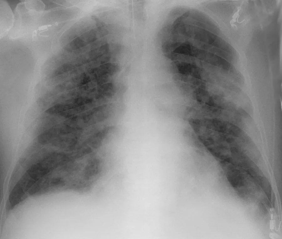
Chest CT scans may be helpful to diagnose COVID‑19 in individuals with a high clinical suspicion of infection but are not recommended for routine screening. Bilateral multilobar ground-glass opacities with a peripheral, asymmetric, and posterior distribution are common in early infection. Subpleural dominance, crazy paving (lobular septal thickening with variable alveolar filling), and consolidation may appear as the disease progresses. Characteristic imaging features on chest radiographs and computed tomography (CT) of people who are symptomatic include asymmetric peripheral ground-glass opacities without pleural effusions.
Many groups have created COVID‑19 datasets that include imagery such as the Italian Radiological Society which has compiled an international online database of imaging findings for confirmed cases. Due to overlap with other infections such as adenovirus, imaging without confirmation by rRT-PCR is of limited specificity in identifying COVID‑19. A large study in China compared chest CT results to PCR and demonstrated that though imaging is less specific for the infection, it is faster and more sensitive.
Coding
In late 2019, the WHO assigned emergency ICD-10 disease codes U07.1 for deaths from lab-confirmed SARS-CoV-2 infection and U07.2 for deaths from clinically or epidemiologically diagnosed COVID‑19 without lab-confirmed SARS-CoV-2 infection.
Pathology
The main pathological findings at autopsy are:
Macroscopy: pericarditis, lung consolidation and pulmonary oedema
Minor serous exudation, minor fibrin exudation
Pulmonary oedema, pneumocyte hyperplasia, large atypical pneumocytes, interstitial inflammation with lymphocytic infiltration and multinucleated giant cell formation
Diffuse alveolar damage (DAD) with diffuse alveolar exudates. DAD is the cause of acute respiratory distress syndrome (ARDS) and severe hypoxaemia.
Organisation of exudates in alveolar cavities and pulmonary interstitial fibrosis
Plasmocytosis in bronchoalveolar lavage (BAL)
Blood and vessels: disseminated intravascular coagulation (DIC); leukoerythroblastic reaction, endotheliitis, hemophagocytosis
Prevention
Preventive measures to reduce the chances of infection include getting vaccinated, staying at home, wearing a mask in public, avoiding crowded places, keeping distance from others, ventilating indoor spaces, managing potential exposure durations, washing hands with soap and water often and for at least 20 seconds, practising good respiratory hygiene, and avoiding touching the eyes, nose, or mouth with unwashed hands.
Those diagnosed with COVID‑19 or who believe they may be infected are advised by the CDC to stay home except to get medical care, call ahead before visiting a healthcare provider, wear a face mask before entering the healthcare provider's office and when in any room or vehicle with another person, cover coughs and sneezes with a tissue, regularly wash hands with soap and water and avoid sharing personal household items.
The first COVID‑19 vaccine was granted regulatory approval on 2 December 2020 by the UK medicines regulator MHRA. It was evaluated for emergency use authorisation (EUA) status by the US FDA, and in several other countries. Initially, the US National Institutes of Health guidelines do not recommend any medication for prevention of COVID‑19, before or after exposure to the SARS-CoV-2 virus, outside the setting of a clinical trial. Without a vaccine, other prophylactic measures, or effective treatments, a key part of managing COVID‑19 is trying to decrease and delay the epidemic peak, known as "flattening the curve". This is done by slowing the infection rate to decrease the risk of health services being overwhelmed, allowing for better treatment of active cases, and delaying additional cases until effective treatments or a vaccine become available.
Indoor ventilation and avoiding crowded indoor spaces
The CDC states that avoiding crowded indoor spaces reduces the risk of COVID-19 infection. When indoors, increasing the rate of air change, decreasing recirculation of air and increasing the use of outdoor air can reduce transmission. The WHO recommends ventilation and air filtration in public spaces to help clear out infectious aerosols.
Exhaled respiratory particles can build-up within enclosed spaces with inadequate ventilation. The risk of COVID‑19 infection increases especially in spaces where people engage in physical exertion or raise their voice (e.g., exercising, shouting, singing) as this increases exhalation of respiratory droplets. Prolonged exposure to these conditions, typically more than 15 minutes, leads to higher risk of infection.
Displacement ventilation with large natural inlets can move stale air directly to the exhaust in laminar flow while significantly reducing the concentration of droplets and particles. Passive ventilation reduces energy consumption and maintenance costs but may lack controllability and heat recovery. Displacement ventilation can also be achieved mechanically with higher energy and maintenance costs. The use of large ducts and openings helps to prevent mixing in closed environments. Recirculation and mixing should be avoided because recirculation prevents dilution of harmful particles and redistributes possibly contaminated air, and mixing increases the concentration and range of infectious particles and keeps larger particles in the air.
Hand-washing and hygiene
Thorough hand hygiene after any cough or sneeze is required. The WHO also recommends that individuals wash hands often with soap and water for at least 20 seconds, especially after going to the toilet or when hands are visibly dirty, before eating and after blowing one's nose. When soap and water are not available, the CDC recommends using an alcohol-based hand sanitiser with at least 60% alcohol. For areas where commercial hand sanitisers are not readily available, the WHO provides two formulations for local production. In these formulations, the antimicrobial activity arises from ethanol or isopropanol. Hydrogen peroxide is used to help eliminate bacterial spores in the alcohol; it is "not an active substance for hand antisepsis". Glycerol is added as a humectant.
Surface cleaning
After being expelled from the body, coronaviruses can survive on surfaces for hours to days. If a person touches the dirty surface, they may deposit the virus at the eyes, nose, or mouth where it can enter the body and cause infection. Evidence indicates that contact with infected surfaces is not the main driver of COVID‑19, leading to recommendations for optimised disinfection procedures to avoid issues such as the increase of antimicrobial resistance through the use of inappropriate cleaning products and processes. Deep cleaning and other surface sanitation has been criticised as hygiene theatre, giving a false sense of security against something primarily spread through the air.
The amount of time that the virus can survive depends significantly on the type of surface, the temperature, and the humidity. Coronaviruses die quickly when exposed to the UV light in sunlight. Like other enveloped viruses, SARS-CoV-2 survives longest when the temperature is at room temperature or lower, and when the relative humidity is low (<50%).
On many surfaces, including glass, some types of plastic, stainless steel, and skin, the virus can remain infective for several days indoors at room temperature, or even about a week under ideal conditions. On some surfaces, including cotton fabric and copper, the virus usually dies after a few hours. The virus dies faster on porous surfaces than on non-porous surfaces due to capillary action within pores and faster aerosol droplet evaporation. However, of the many surfaces tested, two with the longest survival times are N95 respirator masks and surgical masks, both of which are considered porous surfaces.
The CDC says that in most situations, cleaning surfaces with soap or detergent, not disinfecting, is enough to reduce risk of transmission. The CDC recommends that if a COVID‑19 case is suspected or confirmed at a facility such as an office or day care, all areas such as offices, bathrooms, common areas, shared electronic equipment like tablets, touch screens, keyboards, remote controls, and ATMs used by the ill persons should be disinfected. Surfaces may be decontaminated with the following:
Other solutions, such as benzalkonium chloride and chlorhexidine gluconate, are less effective. Ultraviolet germicidal irradiation may also be used, although popular devices require 5–10 min exposure and may deteriorate some materials over time.
Self-isolation
Self-isolation at home has been recommended for those diagnosed with COVID‑19 and those who suspect they have been infected. Health agencies have issued detailed instructions for proper self-isolation. Many governments have mandated or recommended self-quarantine for entire populations. The strongest self-quarantine instructions have been issued to those in high-risk groups. Those who may have been exposed to someone with COVID‑19 and those who have recently travelled to a country or region with the widespread transmission have been advised to self-quarantine for 14 days from the time of last possible exposure.
References
- COVID-19 — Wikipedia, The Free EncyclopediaText from the Wikipedia article "COVID-19" (CC BY-SA 4.0); excerpted. Licence. Retrieved 2026-10-19. Revision used.
Text above is quoted from the listed sources without changes to its meaning. Image credits and licences are shown with each image. Educational information only, not medical advice.
Related conditions
Educational overview, not medical advice. Treatment varies by person and location. Consult a qualified health professional for diagnosis or care. Content and model notes · Privacy information
Social distancing
Social distancing (also known as physical distancing) includes infection control actions intended to slow the spread of the disease by minimising close contact between individuals. Methods include quarantines; travel restrictions; and the closing of schools, workplaces, stadiums, theatres, or shopping centres. Individuals may apply social distancing methods by staying at home, limiting travel, avoiding crowded areas, using no-contact greetings, and physically distancing themselves from others.
In 2020, outbreaks occurred in prisons due to crowding and an inability to enforce adequate social distancing. In the United States, the prisoner population is ageing and many of them are at high risk for poor outcomes from COVID‑19 due to high rates of coexisting heart and lung disease, and poor access to high-quality healthcare.